Central Instrumentation Facility
⇒ Equipments & Facility Access Rules & Guidelines
Name of the Equipment |
Light Emitting Diode Fluorimeter (Uranium Analyser) |
| Model Name | M/s UA-01 |
| Make | Quantalase |
| Specification |
Detector - Photomultiplier tube Excitation source - LEDs Wavelength of emission - As required by the user. Wavelength can be selected between 200nm to 500 nm. Bandwidth of emission - Typically 10-20 nm. Pulse duration - Between 2 to 50 microsecs. To be set by operater. Pulse repetition rate - 1 KHzs. |
|
|
|
| Major Applications | |
| Details of Sample Analysis | Fluorimeters which use banks of pulsed LEDs to excite fluorescence in sample under study. The wavelength, pulse duration and peak power of the LED output can be set to match the excitation requirements of the sample. The fluorescence is detected by a pulsed photomultiplier. Suitable filters after the LEDs and before the photomultiplier tube prevent LED light from reaching the photomultiplier tube directly. The filters can be broad band coloured glass filters or multilayer narrow band filters. The instrument is controlled by a microcontroller which pulses the LEDs and photomultiplier tube. |
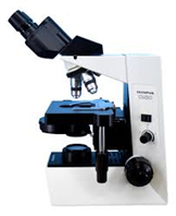
Name of the Equipment |
Phase Contrast Microscope |
| Model Name | Eclips 201, 2012 |
| Make | M/s Nikkon |
| Specification | Episcopic illumination models: ECLIPSE LV150N (manual control types) |
|
|
|
| Major Applications | Phase contrast, by "converting" phase specimens such as living material into amplitude specimens, allowed scientists to see details in unstained and/or living objects with a clarity and resolution never before achieved. |
| Details of Sample Analysis | Unstained specimens that do not absorb light are called phase objects because they slightly alter the phase of the light diffracted by the specimen, usually by retarding such light approximately 1/4 wavelength as compared to the un deviated direct light passing through or around the specimen unaffected. Unfortunately, our eyes as well as camera film, are unable to detect these phase differences. To reiterate, the human eye is sensitive only to the colors of the visible spectrum (variations in light frequency) or to differing levels of light intensity (variations in wave amplitude). In phase specimens, the direct zeroth order light passes through or around the specimen undeviated. However, the light diffracted by the specimen is not reduced in amplitude as it is in a light-absorbing object, but is slowed by the specimen because of the specimen's refractive index or thickness (or both). This diffracted light, lagging behind by approximately 1/4 wavelength, arrives at the image plane out of step (also termed out of phase) with the undeviated light but, in interference, essentially undiminished in intensity. The result is that the image at the eyepiece level is so lacking in contrast as to make the details almost invisible. |
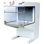
Name of the Equipment |
Laminar Hood |
| Model Name | Vertical Laminar Flow system |
| Make | M/s Alpha Linear |
| Specification |
Pre filter (90% down to 10 micron) HEPA filter (99.97% down to 0.3 micron) Front Side door Polycarbonate/Glass window Manual Operation/ Counterweight Mechanism UV light 5A Power Socket Differential Pressure Gauge |
|
|
|
| Major Applications |
1. Laminar flow cabinets are used in laboratories for contamination sensitive processes like plant tissue culture. 2. Other laboratories processes like media plate preparation and culture of organisms can be performed inside the cabinet. 3. Operations of particle sensitive electronic devices are performed inside the cabinet. |
| Details of Sample Analysis | Microbial Load |
Name of the Equipment |
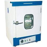
Bacteriological Incubator |
| Model Name | ACM 22062-1 |
| Make | ACMAS, Pvt.Ltd. |
| Specification | An incubator is an insulated and enclosed device used in biological laboratories. It creates an optimum environment which is required for the growth of microorganisms by providing optimum temperature, humidity, and other environmental conditions such as the CO2 and oxygen content inside’s atmosphere. |
|
|
|
| Major Applications | it is used to grow and maintain microbiological cultures or cell cultures. Both bacterial and eukaryotic cell organisms are cultivated by using an incubator. |
| Details of Sample Analysis | Bacterial Culture |
Name of the Equipment |
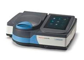
UV- Vis Spectrophotometer |
||||||||||||||||||||||||||||||||||||||
| Model Name | Genesys-50 | ||||||||||||||||||||||||||||||||||||||
| Make | M/s. Thermo Fisher | ||||||||||||||||||||||||||||||||||||||
| Specification |
|
||||||||||||||||||||||||||||||||||||||
|
|
|||||||||||||||||||||||||||||||||||||||
| Major Applications |
-Quantify and qualify DNA, RNA, and protein samples. -Identify sample contaminants and obtain corrected concentration results. |
||||||||||||||||||||||||||||||||||||||
| Details of Sample Analysis | Biological samples | ||||||||||||||||||||||||||||||||||||||
Name of the Equipment |
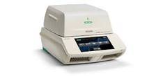
Thermal Cycler PCR |
||||||||||||||||||||||||||
| Model Name | T 100 Thermal cycler | ||||||||||||||||||||||||||
| Make | M/s. Biorad | ||||||||||||||||||||||||||
| Specification |
|
||||||||||||||||||||||||||
|
|
|||||||||||||||||||||||||||
| Major Applications |
Amplification/PCR Cloning Cycle sequencing Gene expression studies Mutagenesis |
||||||||||||||||||||||||||
| Details of Sample Analysis | Biological samples | ||||||||||||||||||||||||||
Name of the Equipment |
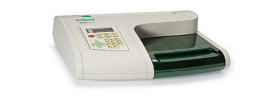
Elisa Reader and Washer |
| Model Name | iMark |
| Make | M/s. Biorad |
| Specification |
Wavelength range - 400–750 nm Photometric range- 0.0–3.5 OD Linearity - ≤1.0% from 0.0–2.0 OD; ≤2.0% from 0.0–3.0 OD Accuracy- ≤1.0% or 0.010 from 0.000–3.000 OD at 490 nm Precision - 1.0% or 0.005 OD from 0.0–2.0 OD; 1.5% from 2.0–3.0 OD Resolution- 0.001 OD Filter wheel capacity- 8 Plate shaking (3 speeds)- Low, mid, high Duration, sec- 0–999 Read time - 6 sec at single wavelength, 10 sec at dual wavelengths Data output - Onboard graphical thermal printer and USB2 interface with PC or Mac data stations Data storage - Calender/clock functions; 64 assay protocols Multilanguage support - 4 languages, LCD indication supported; printout report supported Dimensions (W x D x H), cm (in)- 34.6 x 37.7 x 16.4 (13.6 x 14.8 x 6.5) Weight, kg (lb)- 5.5 (12) |
|
|
|
| Major Applications | Immunological Studies |
| Details of Sample Analysis | Biological samples |
Name of the Equipment |
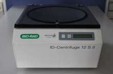
Refrigerated Centrifuge |
| Model Name | Sorvall legend micro.17R |
| Make | M/s. Biorad |
| Specification |
ID-Centrifuge 12 S II is a silent centrifuge for up to 12 ID-Cards with automatic balance control. The centrifugation time is 10 minutes. Speed: 1030 rpm (85 g) Dimensions (w/h/d): 30 cm / 18 cm / 36 cm Weight: 6.2 kg Power requirements: 100-240 V / 50-60 Hz |
|
|
|
| Major Applications |
• Separation of mixtures with close densities. • Separate immiscible liquids. • Sediment suspended solids. • Separation of blood. • Separation insoluble particles (e.g. insoluble proteins in a protein solution) • Isotope Separation. |
| Details of Sample Analysis | Biological samples |
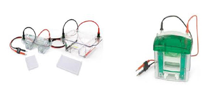
Name of the Equipment |
Horizontal and Vertical Electrophoresis unit |
| Model Name | Mini-Protrean Tetra cell and Mini-Sub cell GT Respectively |
| Make | M/s. Biorad |
| Specification |
Number of gels 1–4 Precast gels Ready Gel precast gels Handcast gels Cast using Mini-PROTEAN spacer plates Gel size (W x L) Precast: 8.6 x 6.8 cm Handcast: 8.3 x 7.3 cm Glass plate size (W x L) Short plate 10.1 x 7.3 cm Spacer plate 10.1 x 8.2 cm Total buffer volume for 2 gels 700 ml Total buffer volume for 4 gels 1,000 ml Typical run times for SDS-PAGE 35–45 min (at 200 V constant) Recommended power supply PowerPac Basic or PowerPac HC (High Current) Dimensions (W x L x H) 12 x 16 x 18 cm |
|
|
|
| Major Applications | horizontal gel electrophoresis is an ideal choice for DNA and RNA separation, while vertical systems are ideal for proteins |
| Details of Sample Analysis | DNA, RNA and Proteins |